Anatomy of the Human Heart
Welcome to United Medical Education. Today we will be discussing the anatomy and function of the human heart. The human heart is a pump made primarily of cardiac muscle that constantly circulates blood throughout the human body. By so doing, the human heart provides nutrients and oxygen to organs, including itself. The circulated blood also allows for the removal of carbon dioxide and other cellular waste. Even though the heart functions as a powerful pump, it is only roughly the size of a human fist. The average male human heart is approximately 10 to 12 ounces while the average heart of a woman is approximately 8 to 10 ounces.
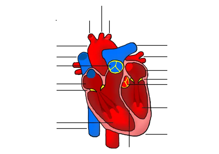
The human heart is made up of four chambers and four valves. These chambers and valves are made up of cardiac muscle and connective tissue. There are two upper chambers called the right atrium and the left atrium. These two smaller chambers feed the larger two lower chambers called the right ventricle and the left ventricle. The right atrium and the right ventricle together make up the right side of the heart. The right atrium and the right ventricle are separated by a valve called the atrioventricular (AV) or tricuspid valve. The semi-lunar or pulmonic valve separates the right ventricle from the pulmonary artery in the pulmonary circuit. The right side of the heart deals with deoxygenated blood.
The left atrium and the left ventricle together make up the left side of the heart. They are separated also by an atrioventricular (AV) valve but this one is called the mitral valve. There is also the aortic valve that separates the left ventricle from the systemic circuit. The left side of the heart deals with oxygenated blood. There are also what’s called chordae tendinae that attach to each of the heart valves and assure the correct opening and closing of the valves as appropriate pressure builds within the chambers.
The heart’s wall is made up of three distinct layers. The inner layer is referred to as the endocardium and is what makes contact with the circulating blood. The middle layer is referred to as the myocardium and is primarily made up of the cardiac muscle that contracts the heart. The outer layer is the epicardium and makes up the inner wall of a structure that is called the pericardium.
Surrounding the heart there is a layer called the pericardium. The pericardium protects, supports, and lubricates the outside of the heart to prevent friction and lower energy expenditure. The pericardium has two layers. The inner layer of the pericardium is referred to as the serous pericardium. The outer layer is referred to as the parietal pericardium. Between the two layers of the pericardium lies the lubricating fluid called pericardial fluid.
Function of the Human Heart
The function of the human heart begins with an electrical impulse that originates from a location called the atrioventricular (AV) node. the AV node is located in the right atrium and coordinates the contraction of the chambers in the heart muscle. The human heart pumps blood through two separate circuits in the body. First, deoxygenated blood enters the heart through the vena cava and into a chamber called the right atrium. Deoxygenated blood is then pumped through the tricuspid valve to a larger and more powerful chamber called the right ventricle. The right ventricle pumps the deoxygenated blood through the pulmonic valve to the pulmonary circuit. In the pulmonary circuit the deoxygenated blood first passes through the pulmonary arteries to be oxygenated by the lungs. After the deoxygenated blood passes through the lungs vasculature and becomes oxygenated, it returns to the heart as oxygenated blood by means of the pulmonary veins.
Returning to the heart from the pulmonary veins, the blood enters the chamber called the left atrium. The oxygenated blood is then pumped through the mitral valve to a larger and more powerful chamber called the left ventricle. The left ventricle then pumps the oxygenated blood through the aortic valve sending it to the systemic circuit, beginning with the ascending aorta.
The contraction of the human heart is off-set between the atria and the ventricles. First the atria contract and then the ventricles contract shortly after. The correct contraction of the atria is referred to as S1, while the correct contraction of ventricles is referred to as S2. Correct rhythm and contraction of the chambers is necessary for the heart to function efficiently.
Heart Disease
Heart disease continues to be the leading cause of death in the United States. Currently, heart disease costs us $363 billion every year and claims a life in the United States every 36 seconds. As a result, understanding and treating cardiovascular disease, such as heart failure, is one of the greatest medical undertakings of our time. While new technology and treatments are making progress in the fight against heart disease it still remains our greatest threat in healthcare.